
FIGURE
Fig. S4
Fig. S4
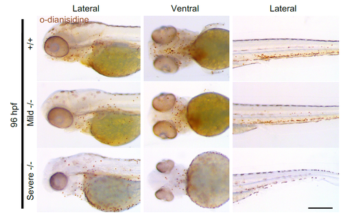
O-Dianisidine staining demonstrates presence of hemoglobinized blood in severe homozygous pdgfra larvae. Representative bright field images of OD-stained larvae indicating the presence of hemoglobinized blood in +/+, mild -/- and severe -/- larvae at 96 hpf. Lateral and ventral views are shown, with the fish cranium on the left. Scale bar: 250 μm. |
Expression Data
Expression Detail
Antibody Labeling
Phenotype Data
Phenotype Detail
Acknowledgments
This image is the copyrighted work of the attributed author or publisher, and
ZFIN has permission only to display this image to its users.
Additional permissions should be obtained from the applicable author or publisher of the image.
Full text @ Biol. Open