|
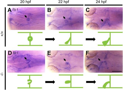
Migration of endocardial precursors to the midline is abnormal in pdgfra mutants. Representative photomicrographs of whole-mount in situ hybridization using fetal liver kinase 1 (flk1) riboprobe. In +/+ embryos, endocardial precursors migrate and fuse at the midline forming a ring-like structure at 20 hpf (A), followed by elongation and leftward movement at 22 (B) and 24 (C) hpf. In −/− mutants, endocardial precursors form a V-like structure at 20 hpf (D). Endocardial cells reach the midline and begin to elongate at 22 hpf (E). Elongation continues with abnormal leftward movement at 24 hpf (F). Arrows point to endocardium. Dorsal views are shown, with the fish cranium on the left. An illustration of the developing heart is shown at the bottom of each figure. Scale bar: 100 μm.
|