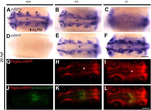
Pdgfra is expressed in the midline during cardiac assembly. Representative photomicrographs of whole-mount in situ hybridization using pdgfra and mRFP riboprobes at 20 hpf (A-F). In +/+ (A) and +/− (B) embryos expression of pdgfra is observed in different tissues, including the developing cranial ganglia (arrow head), anterior lateral plate mesoderm (ALPM) (arrow), optic cup (cross), and the midline (asterisk, also in panels E and F). No specific pdgfra expression is detected in −/− embryos (C). Expression of mRFP is not detected in +/+ embryos (D). On the other hand, mRFP expression in +/− (E) and −/− (F) embryos is similar to pdgfra expression. Representative images of 20 hpf live GBT1300 siblings carrying a Tg(cmlc2:EGFP) transgene (G-L). No Pdgfra-mRFP expression is observed in +/+ embryos (G). On the other hand, +/− (H) and −/− (I) siblings show Pdgfra-mRFP expression similar to the mRNA expression data, including midline expression where cardiac assembly occurs (asterisk). Complete cardiac fusion is observed in +/+ (J) and +/− (K) embryos, while homozygous mutants demonstrated cardiac fusion only at the posterior end (L). Dorsal views are shown, with the fish cranium on the left. Scale bar: 75 μm (A-F) and 250 μm (G-L).
|
