
FIGURE
Fig. 3
Fig. 3
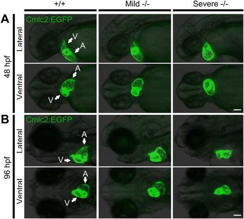
Pdgfra homozygous mutants exhibit abnormal cardiac phenotypes. An incross of the pdgfra gene-trapped strain carrying a Tg(cmlc2:EGFP) transgene was used to investigate the cardiac phenotype. (A) At 48 hpf, mild and severe −/− mutants present with abnormal cardiac looping; severe −/− embryos also develop an enlarged atrium and pericardial edema. (B) At 96 hpf, the hearts of mild −/− mutants remain incompletely looped, while severe −/− mutants present with collapsed chambers and pericardial edema. Arrows pointing at ventricle (V) and atrium (A). The fish cranium is on the left. Scale bar: 100 μm. |
Expression Data
| Gene: | |
|---|---|
| Fish: | |
| Anatomical Term: | |
| Stage Range: | Long-pec to Day 4 |
Expression Detail
Antibody Labeling
Phenotype Data
| Fish: | |
|---|---|
| Observed In: | |
| Stage Range: | Long-pec to Day 4 |
Phenotype Detail
Acknowledgments
This image is the copyrighted work of the attributed author or publisher, and
ZFIN has permission only to display this image to its users.
Additional permissions should be obtained from the applicable author or publisher of the image.
Full text @ Biol. Open