
Fig. 6
- ID
- ZDB-FIG-170424-9
- Publication
- Schmitner et al., 2017 - ptf1a+ , ela3l- cells are developmentally maintained progenitors for exocrine regeneration following extreme loss of acinar cells in zebrafish larvae.
- Other Figures
- All Figure Page
- Back to All Figure Page
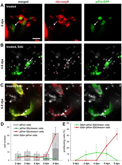
Ptf1a+ cells as a source for exocrine cell regeneration. (A-C) Confocal images of the pancreatic head region of EdU-labeled Tg(ela:casp8; ptf1a:eGFP) at 0 dpa (A), 6 dpa (B) and 8 dpa (C) following a 7-9 dpf ablation treatment showing that during regeneration, ptf1a+, ela3l− cells (white arrows) rarely proliferate and differentiated exocrine cells reconstitute the exocrine pancreas. (D) Absolute numbers of all ptf1a+ cells in the pancreatic head region, proliferating ptf1a+ cells and GFP+, E2Crimson− and GFP+, E2Crimson+ cells. (E) Relative amount of EdU+, ptf1a+, E2Crimson− and EdU+, ptf1a+, E2Crimson+ cells. Mean+s.e.m. Scale bar: 20 µm. |