
Fig. 2
- ID
- ZDB-FIG-170424-5
- Publication
- Schmitner et al., 2017 - ptf1a+ , ela3l- cells are developmentally maintained progenitors for exocrine regeneration following extreme loss of acinar cells in zebrafish larvae.
- Other Figures
- All Figure Page
- Back to All Figure Page
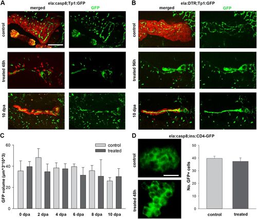
Ablation of exocrine cells does not affect neighboring NRCs and β-cells. (A,B) Confocal image projections of ela:casp8;Tp1:eGFP (A) and ela:DTR;Tp1:eGFP (B) in untreated control animals (7 dpf for ela:casp8 and 9 dpf for ela:DTR), animals directly after treatment and 10 dpf animals. Note that newly established exocrine cells are found next to NRCs. (C) Volume of pancreatic NRCs, determined by GFP expression, is not significantly changed after ablation and during regeneration of exocrine cells in ela:casp8 embryos (n>5 larva for each time point). (D) β-cells labeled using Tg(ins:CD4-eGFP). The number of β-cells was unaffected by treatment with dimerizer (n=6). Values are mean+s.e.m. Scale bars: 100 µm (A,B) and 20 µm (D). |