
Fig. 3
- ID
- ZDB-FIG-170424-6
- Publication
- Schmitner et al., 2017 - ptf1a+ , ela3l- cells are developmentally maintained progenitors for exocrine regeneration following extreme loss of acinar cells in zebrafish larvae.
- Other Figures
- All Figure Page
- Back to All Figure Page
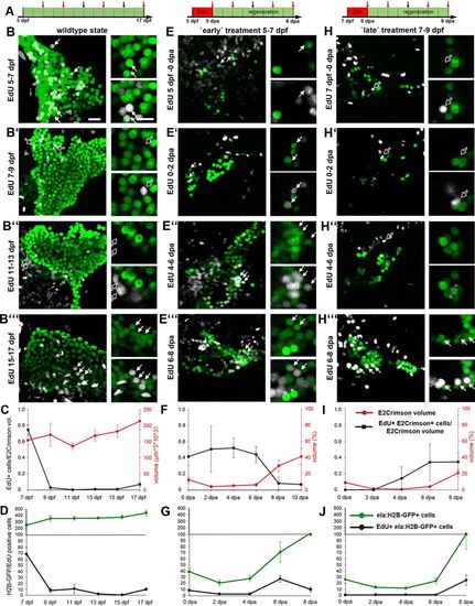
Dynamics of exocrine development and regeneration. (A) Time scheme of regeneration experiments indicating phase with Dim treatment (in red) and time points of fixation (arrows). (B,E,H) Confocal images of Tg(ela:casp8;ela:H2B-GFP) animals at different stages of larval development (stages as indicated by red arrows in A). All larvae were treated with EdU for 48 h before fixation (GFP signals in green, EdU signals in white, smaller white dots correspond to E2Crimson background). Images cover the pancreatic head region of untreated (B-B′″) animals and of larvae treated with 5 µM Dim either from 5 to 7 dpf (E-E″′, ‘early’ treatment) or from 7 to 9 dpf (H-H′″, ‘late’ treatment). Shown are projections and selections of single plane images (smaller images to the right). Note that ‘late’ treatment larva between 0-6 dpa lack EdU+ GFP+ cells (white arrows) and that EdU+ signals in 9 dpf and 13 dpf control animals (B′,B″) and in 0-6 dpa late treatment larvae (H-H″) localized to GFP-negative nuclei (black arrows). (C,D,F,G,I,J) Data quantification (n>5 larva for each time point; mean+s.e.m.) using volumetric measurements (C,F,I) and cell counts (D,G,J: absolute numbers of nuclei per stack) for control larva (C,D), early treatment larvae (F,G) and late treatment larvae (I,J). Red line in C shows the absolute E2Crimson volume (in red), whereas in F and I, it shows the relative volume compared with the wild-type situation as shown in C. Note that different samples were analyzed in E-G and H-J, and that the higher proliferation rate in 11 dpf control animals shown in D compared with C result from only two larvae with 10 and 40 EdU+ cells (see Table S1). Scale bars: 20 µm. |