
FIGURE
Fig. S5
- ID
- ZDB-FIG-170424-14
- Publication
- Schmitner et al., 2017 - ptf1a+ , ela3l- cells are developmentally maintained progenitors for exocrine regeneration following extreme loss of acinar cells in zebrafish larvae.
- Other Figures
- All Figure Page
- Back to All Figure Page
Fig. S5
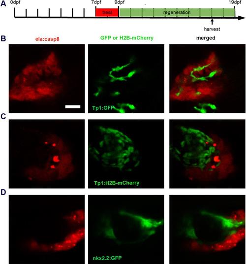
Duct cells are not involved in exocrine cell regeneration. A: timeline indicating time of treatment and harvesting of larvae. Confocal images of regeneration of exocrine cells at 8 dpa in tg(Tp1:GFP) (B), tg(Tp1:H2B-mCherry) (C) and tg(nkx2.2a:GFP) (D) background showing newly formed exocrine cells adjacent to the duct and no overlapping fluorophore expression. Scale bar: 20μm. |
Expression Data
Expression Detail
Antibody Labeling
Phenotype Data
Phenotype Detail
Acknowledgments
This image is the copyrighted work of the attributed author or publisher, and
ZFIN has permission only to display this image to its users.
Additional permissions should be obtained from the applicable author or publisher of the image.
Full text @ Dis. Model. Mech.