
FIGURE
Fig. 2
- ID
- ZDB-FIG-170413-30
- Publication
- Carroll et al., 2014 - Estrogen defines the dorsal-ventral limit of VEGF regulation to specify the location of the hemogenic endothelial niche
- Other Figures
- All Figure Page
- Back to All Figure Page
Fig. 2
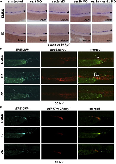
ERE:GFP Embryos Display Estrogen Activity in the Developing AGM (A) MO knockdown showed that the effect of E2 on HSPCs is mediated through esr2a+b; esr1-MO had no impact on runx1/cmyb (n ≥ 30). (B) ERE:GFP embryos show low expression in the trunk vasculature, labeled by lmo2:dsRed; ERE activity expands following E2 exposure and is alleviated by treatment with ZK164015 (n ≥ 20). (C) ERE:GFP embryos exhibit activity in the developing mesonephros, as indicated by cdh17:mCherry at 48 hpf, which responds to altered E2 signaling (n ≥ 20). |
Expression Data
| Gene: | |
|---|---|
| Fish: | |
| Condition: | |
| Knockdown Reagents: | |
| Anatomical Term: | |
| Stage: | Prim-25 |
Expression Detail
Antibody Labeling
Phenotype Data
| Fish: | |
|---|---|
| Condition: | |
| Knockdown Reagents: | |
| Observed In: | |
| Stage: | Prim-25 |
Phenotype Detail
Acknowledgments
This image is the copyrighted work of the attributed author or publisher, and
ZFIN has permission only to display this image to its users.
Additional permissions should be obtained from the applicable author or publisher of the image.
Reprinted from Developmental Cell, 29, Carroll, K.J., Esain, V., Garnaas, M.K., Cortes, M., Dovey, M.C., Nissim, S., Frechette, G.M., Liu, S.Y., Kwan, W., Cutting, C.C., Harris, J.M., Gorelick, D.A., Halpern, M.E., Lawson, N.D., Goessling, W., North, T.E., Estrogen defines the dorsal-ventral limit of VEGF regulation to specify the location of the hemogenic endothelial niche, 437-53, Copyright (2014) with permission from Elsevier. Full text @ Dev. Cell