
Fig. 4
- ID
- ZDB-FIG-170413-32
- Publication
- Carroll et al., 2014 - Estrogen defines the dorsal-ventral limit of VEGF regulation to specify the location of the hemogenic endothelial niche
- Other Figures
- All Figure Page
- Back to All Figure Page
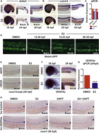
Exogenous E2 Signaling Disrupts VEGF/NOTCH Cascades in the AGM (A) E2 exposure had no impact on deltaC expression by WISH at 18 hpf but strongly reduced arterial expression at 24 and 36 hpf (n > 30/treatment); an arrowhead indicates artery. (B) Expression of notch3 was similarly regulated by E2 at 18, 24, and 36 hpf (n > 30/treatment); an arrowhead indicates artery. (C) qPCR confirmed E2-mediated reductions in deltaC and notch3 expression at 36 hpf (mean of triplicate experiments ± SEM; one-tailed t test deltaC, notch3∗p < 0.05). (D) Decreased Notch:GFP was observed in the artery (white bracket indicates artery walls) after E2 treatment from 12–36 or 12–23 hpf; no change occurred with exposure from 26–36 hpf (n ≥ 52). (E) NICD induction increased expression of runx1/cmyb and rescued HSPCs in E2-treated embryos (n ≥ 31/treatment). (F) Expression of VEGFAa was strongly decreased in zebrafish following E2 treatment at both 18 and 24 hpf (n ≥ 25/treatment); an arrow points to somite staining. (G) qPCR confirmed that expression of VEGFAa was decreased at 24 hpf (mean of triplicate experiments ± SEM; one-tailed t test ∗∗∗p < 0.001). (H) Induction of VEGFAa increased runx1 expression and rescued the effect of E2 exposure; treatment with DAPT decreased expression of runx1 and blocked the VEGF-mediated rescue of HSPCs (n ≥ 20/treatment); an arrowhead indicates artery. |
Reprinted from Developmental Cell, 29, Carroll, K.J., Esain, V., Garnaas, M.K., Cortes, M., Dovey, M.C., Nissim, S., Frechette, G.M., Liu, S.Y., Kwan, W., Cutting, C.C., Harris, J.M., Gorelick, D.A., Halpern, M.E., Lawson, N.D., Goessling, W., North, T.E., Estrogen defines the dorsal-ventral limit of VEGF regulation to specify the location of the hemogenic endothelial niche, 437-53, Copyright (2014) with permission from Elsevier. Full text @ Dev. Cell