
FIGURE
Fig. 3
Fig. 3
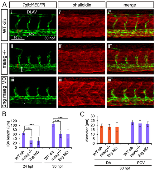
maeg loss of function results in the blood vessel morphogenesis defects in zebrafish embryos. A. Confocal imaging analysis of trunk vascular and somital morphology in WT, maeg−/− and maeg morphants Tg(kdrl:EGFP) embryos at 30 hpf. Red and white square brackets indicate the lumen of the DA and PCV, respectively. DA, dorsal aorta; PCV, posterior cardinal vein; and DLAV, dorsal longitudinal anastomotic vessel. B. The statistics of ISV length in WT, maeg−/− and maeg morphants. One-Way ANOVA; ***,P<0.001. C. The statistics of DA and PCV lumen size at 30 hpf. Error bars indicate stdev. |
Expression Data
| Gene: | |
|---|---|
| Fish: | |
| Knockdown Reagent: | |
| Anatomical Terms: | |
| Stage: | Prim-15 |
Expression Detail
Antibody Labeling
Phenotype Data
| Fish: | |
|---|---|
| Knockdown Reagent: | |
| Observed In: | |
| Stage Range: | Prim-5 to Prim-15 |
Phenotype Detail
Acknowledgments
This image is the copyrighted work of the attributed author or publisher, and
ZFIN has permission only to display this image to its users.
Additional permissions should be obtained from the applicable author or publisher of the image.
Full text @ Oncotarget