
FIGURE
Fig. S2
Fig. S2
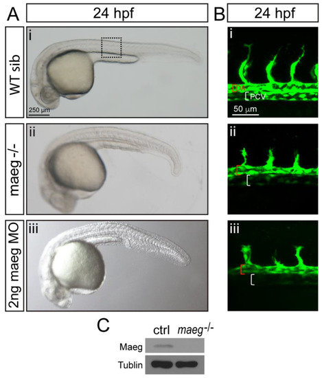
maeg loss of function results in the blood vessel morphogenesis defects in zebrafish embryos. A. Zebrafish embryos of WT, maeg−/− and maeg morphants at 24 hpf imaged in bright field. Square in dash line indicates the confocal imaging region of panel B. B. Confocal imaging analysis of trunk vascular morphology in WT, maeg−/− and maeg morphants Tg(kdrl:EGFP) embryos at 24 hpf. Red and white square brackets indicate the lumen of the DA and PCV, respectively. DA, dorsal aorta; PCV, posterior cardinal vein. C. Western blot analysis of Maeg expression in 24hpf control and mutants whole embryos. |
Expression Data
Expression Detail
Antibody Labeling
Phenotype Data
Phenotype Detail
Acknowledgments
This image is the copyrighted work of the attributed author or publisher, and
ZFIN has permission only to display this image to its users.
Additional permissions should be obtained from the applicable author or publisher of the image.
Full text @ Oncotarget