
Fig. 4
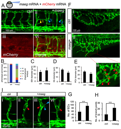
Maeg overexpression causes excessive branching. A. Confocal imaging analysis of trunk vascular morphology in control and maeg/mCherry mRNA mixture injected Tg(kdrl:EGFP) embryos at 48 hpf. Yellow arrowhead indicates the aberrant vessel connected two adjacent ISVs. Blue arrowheads indicate Y-shaped ISVs. B. The statistics of hyperbranching sprouts in maeg up-regulated embryos. C-E. The statistics of ISV, DA and PCV lumen size at 48 hpf. Error bars indicate s.e.m. F. Morphology of subintestinal vessel (SIVs) in 72 hpf Tg(fli1a:EGFP) embryos injected with control mRNA or maeg mRNA. G. The statistics of branch point number. Student’s t-test; ***,P<0.001. H. Quantification of ECs nuclei number in SIVs. Student’s t-test; ***,P<0.001. I. Confocal imaging analysis of ISVs morphology in control and maeg mRNA injected Tg(kdrl:EGFP) embryos at 48 hpf. Red arrowheads indicate knot-like structures. Blue arrowheads indicate angiogenic sprouts. |