
Fig. S5
- ID
- ZDB-FIG-170209-56
- Publication
- Noack Watt et al., 2016 - The Roles of RNA Polymerase I and III Subunits Polr1c and Polr1d in Craniofacial Development and in Zebrafish Models of Treacher Collins Syndrome
- Other Figures
- All Figure Page
- Back to All Figure Page
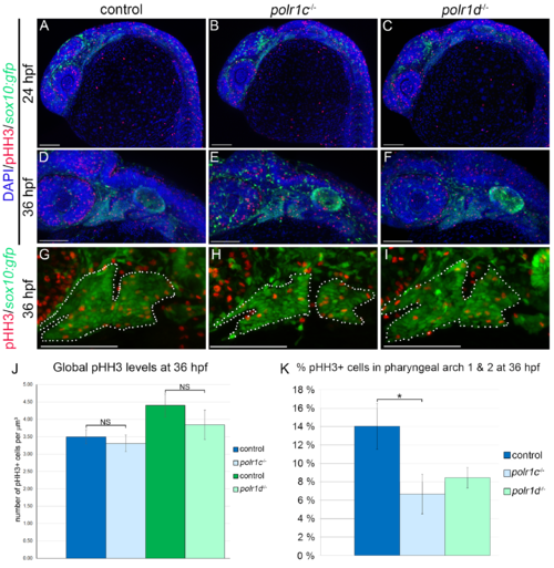
Proliferation within pharyngeal arches 1 & 2 is reduced. (A-C) similar levels of pHH3 staining are present in control, polr1c-/- and polr1d-/- embryos at 24 hpf. (D-F) Proliferation at 36 hpf also occurs globally at broadly similar levels in controls and mutant embryos, but there differences in the number of pHH3+ cells within pharyngeal arches 1 and 2. (G-I) Magnified views of pharyngeal arches 1 and 2 (outlined). (J, K) Quantification of pHH3+ labeled cells illustrating no global overall decrease in mutants embryos compared to controls, but a significant decrease in the percentage of pHH3+ cells in pharyngeal arches 1 and 2 in polr1c mutant embryos. polr1d mutant embryos showed a similar level of proliferation as polr1c mutants. Scale bar = 100 μm. * = p < 0.01 and error bars represent 95% confidence intervals. |
| Fish: | |
|---|---|
| Observed In: | |
| Stage: | Prim-5 |