
FIGURE
Fig. 6 S1
- ID
- ZDB-FIG-161103-28
- Publication
- Perez-Camps et al., 2016 - Quantitative imaging reveals real-time Pou5f3-Nanog complexes driving dorsoventral mesendoderm patterning in zebrafish
- Other Figures
- All Figure Page
- Back to All Figure Page
Fig. 6 S1
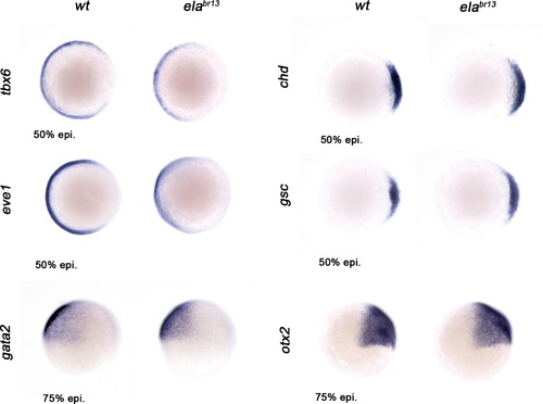
Ela/Aplnr pathway refines ventrolateral Bmp signaling. Expression of the ventrolateral mesoderm markers, tbx6 and eve1, was slightly down-regulated in elabr13 relative to wt embryos at 50% epiboly. Chd expression in the dorsal margin and gsc in the prospective shield does not change in elabr13ascompared with wt embryos at 50% epiboly. Ectoderm markers, gata2 (non-neural ectoderm) and otx2 (forebrain-midbrain), remained similar in elabr13 relative to wt embryos at 75% epiboly. |
Expression Data
Expression Detail
Antibody Labeling
Phenotype Data
Phenotype Detail
Acknowledgments
This image is the copyrighted work of the attributed author or publisher, and
ZFIN has permission only to display this image to its users.
Additional permissions should be obtained from the applicable author or publisher of the image.
Full text @ Elife