
Fig. 5 S1
- ID
- ZDB-FIG-161103-26
- Publication
- Perez-Camps et al., 2016 - Quantitative imaging reveals real-time Pou5f3-Nanog complexes driving dorsoventral mesendoderm patterning in zebrafish
- Other Figures
- All Figure Page
- Back to All Figure Page
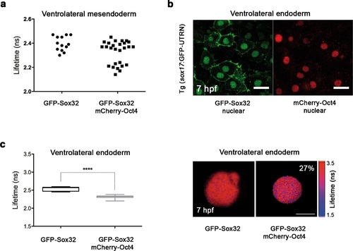
Sox32 binds Oct4 in ventrolateral endoderm. Lifetime values of GFP-Sox32 lifetime alone and co-expressed with mCherry-Oct4 in the nuclei of individual cells from the row next to the margin in ventrolateral mesendoderm at 50% epiboly (5.7 hpf) before involution starts. Those showing a decrease in the lifetime are endoderm precursors and those with similar lifetime are mesoderm precursors. (b) GFP-Sox32 and mCherry-Oct4 co-expressed in Tg(sox17:GFP-UTRN) embryos at 60% epiboly (7 hpf). Scale bar: 20 µm. (c) Lifetime values and FLIM images of GFP-Sox32 lifetime alone and co-expressed with mCherry-Oct4 in the nuclei of individual cells in ventrolateral endoderm at 60% epiboly (7 hpf). The percentage of binding is indicated at the top right corner of the FLIM image. Values represent the median and quartile ranges of data from three independent experiments (n = 20-30 cell nuclei from 10 embryos; ****p<0.0001). Scale bar: 5 µm. |