
Fig. 1 S1
- ID
- ZDB-FIG-161103-17
- Publication
- Perez-Camps et al., 2016 - Quantitative imaging reveals real-time Pou5f3-Nanog complexes driving dorsoventral mesendoderm patterning in zebrafish
- Other Figures
- All Figure Page
- Back to All Figure Page
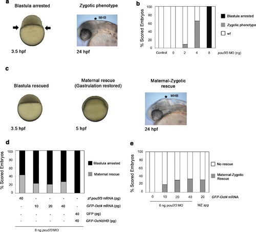
GFP-Oct4 rescues zebrafish Pou5f3 function. (a) Phenotypes of pou5f3 knockdown using morpholino (MO) antisense. Arrows show a constriction in the interface between the yolk and the blastoderm, prohibiting gastrulation in embryos arrested at the blastula stage after MO injection. Embryos not arrested show zygotic phenotype: they do not develop the mid-hindbrain boundary (*MHB) at the 24-hpf stage. (b) Relative percentages of different phenotypes according to the dose of pou5f3 MO injected. Phenotypes are expressed as a percent of whole (n = 150-400 embryos per condition). As a control, a mismatch MO sequence was injected resulting in 100% of embryos with wt phenotype. 100% of embryos were arrested at the blastula stage using 8 ng of MO. Lower-dosed 4-ng and 2-ng embryos continued through gastrulation but did not develop the MHB, according to the percentages shown. (c) Phenotypes of rescued embryos. Rescue of maternal pou5f3 function, as shown by restoring normal gastrulation. Rescue of maternal-zygotic pou5f3 function, as shown by MHB formation at 24 hpf. (d and e) Relative percentages of (d) maternal and (e) maternal-zygotic rescue according to different amounts of injected mRNA. Phenotypes of the rescued embryos are expressed as a percent of whole (n = 170-400 embryos per condition). |