
Fig. 3
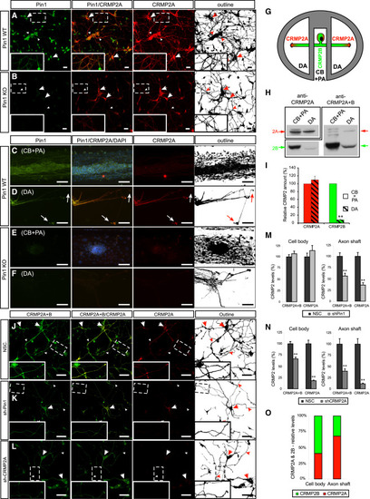
Pin1 Stabilizes CRMP2A Selectively in the Distal Neurites of Primary Neurons (A and B) Pin1 KO reduces CRMP2A selectively in the distal neurites of primary neurons. CRMP2A (red) and Pin1 (green) immunostaining in Pin1 WT and KO primary cortical neurons at 3 DIV. (A) CRMP2A is expressed strongly in the soma (arrows) as well in the neurites (arrowheads) and co-localizes with Pin1 in the neurites (insets). In the Pin1 KO neurons (B), CRMP2A levels are lower in the neurites (arrowheads), but its levels in the somas are comparable to WT (arrows). (C-F) Pin1 KO reduces CRMP2A in axons. Using rat Pin1 WT DRG compartmental cultures, Pin1 was detected both in the cell body and proximal axon (CB + PA) compartment (C) and in distal axons (DA) (D) by double immunostaining, while higher expression of CRMP2A (co-localizing with Pin1) was detected in DA close to growth cones (D, arrows) when compared to the CB + PA (C). In the Pin1 KO DRG neurons, CRMP2A expression in the DA (F), but not in the CB + PA region (E), is significantly reduced. (G-I) The relative level of CRMP2A increases in distal axon region. Lysates collected from DA and CB + PA compartments of rat primary DRG compartment cultures (G) were analyzed by western blotting using CRMP2A and total CRMP2A+B antibodies (H). Semiquantitative analysis of CRMP2A and CRMP2B levels shows significant reduction of CRMP2B levels in distal axons but no significant reduction of CRMP2A levels (I). (J-L) Pin1 KD reduces CRMP2A and total CRMP2 selectively in neurites. Pin1 WT primary cortical neurons were infected with non-silencing (NSC), Sh-Pin1, or Sh-CRMP2A lentiviruses and immunostained for CRMP2A (red) or total CRMP2 (CRMP2A+B) (green). In NSC neurons, high levels of CRMP2A and total CRMP2 (CRMP2A+B) were detected in both the neurites (arrowhead) and the soma (arrow) (J). Pin1 KD significantly reduced CRMP2A and total CRMP2 levels in neurites (arrowhead), but not in cell bodies (arrow) (K). CRMP2A KD significantly decreases CRMP2A and total CRMP2 levels in neurites (arrowhead) as well as cell bodies (arrows) (L). (M and N) Quantification of total CRMP2 and CRMP2A levels in Pin1 KD (shPin1) and non-silencing shRNA (NSC) control neurons in the neuronal cell body and in the axon shafts (M). (O) Quantification of total CRMP2B and CRMP2A levels in CRMP2A KD (shCRMP2A) and control (NSC) neurons in the neuronal cell body and axon shafts. Relative distribution of CRMP2A versus CRMP2B in the cell body and distal axons calculated from (N). Scale bars represent 20 µm (A and B) and 50 µm (C-F and J-L). Data are means ± SEM; p < 0.0001. See also Figure S2. |
| Fish: | |
|---|---|
| Knockdown Reagents: | |
| Observed In: | |
| Stage: | Prim-5 |