
Fig. S2
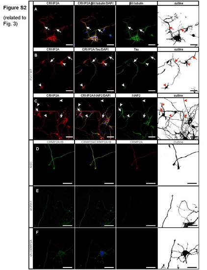
Knockdown of Pin1 or CRMP2A leads to reduction of total CRMP2 level in the vicinity of growth cones. Related to Figure 3 (A) CRMP2A is strongly expressed in primary cortical neurons, as detected by neuron marker βIII tubulin (arrows). (B) Strongest CRMP2A staining is present in distal axons (arrows), as identified by co-immunostaining with tau. CRMP2A is also present in neuronal cell bodies (arrowheads). (C) CRMP2A is present also in dendrites of primary cortical neurons (arrowheads), as identified by the dendritic marker MAP2, even though its level seems lower when compared to CRMP2A level in distal axons (arrows). Knockdown of Pin1 (E) or CRMP2A (F) in the Pin1 WT primary cortical neurons significantly reduces level of CRMP2A as well as total CRMP2 (CRMP2A+B) in the vicinity of growth cones, when compared to control cortical neurons infected with a non-silencing lentiviral vector (D). (Scale bars A – C 50 µm, D – F 20 µm). |