
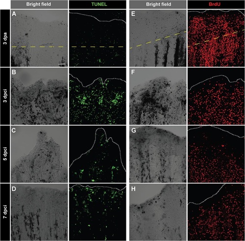
Fig. 6
Enhanced cell proliferation and upregulation of tissue remodelling protein during regeneration of cryoinjured fins. (A-D) TUNEL labelling (green) of whole-mount fins at 3dpa (A) and at different time points after cryoinjury (B-D). At 3dpa (A), TUNEL staining is nearly absent. At 3dpci (B), 5dpci (C) and 7dpci (D), tissue debris (dark structures in bright-field images) are associated with TUNEL-positive cells. (E-H) Immunodetection of BrdU (red) in whole-mount caudal fins at 3dpa (E) and at different time points after cryoinjury (F-H). As compared to 3dpa (E), BrdU-incorporation is lower in cryoinjured fins at 3dpci (F), especially at the position of necrotic cells (dark regions in the bright-field). Cell proliferation becomes more abundant at 5dpci (G) and 7dpci (H). The dashed yellow line indicates the plane of amputation. The edge of the fin is indicated with a white dotted line. N=5. Scale bar=100µm. |