
Fig. 4
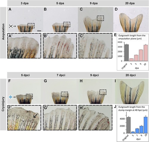
Truncation of the damaged fin tissue is followed by resumed regeneration. (A-D) Time-lapse imaging of fins after amputation during the outgrowth-formation, with boxed areas magnified in A′-C′. (F-I) Time-lapse imaging of fins after cryoinjury during the regenerative phase, with boxed areas magnified in F′-H′. Despite a delay of fin loss and partial damage of the stump, the regenerate reproduces a normal shape of the original fin within 20days. (F2) Arrow indicates a broken bone. (E,J) Quantification of the fin regeneration after amputation (E) and cryoinjury (J). The length of the 3 longest lateral rays was measured from the stump margin after fin loss to the distal tip of the regenerate at different time-points. Error bars represent s.e.m., N=4 fins. Scale bar in A=1mm. |