
Fig. 3
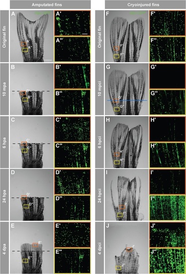
Accumulation of neutrophils in the damaged tissue indicates an acute inflammatory response after cryoinjury. In-vivo visualization of neutrophils in Tg(mpx:GFP) fish. (A-J) Time-lapse bright-field images of the same fins after amputation (A-E) and cryoinjury (F-J). Frames indicate the regions selected for fluorescence imaging of GFP-positive neutrophils, depicted in A′-E′′,F′-J′′ Middle region of the fin (orange box) at the level of the amputation plane (dashed line) and cryoinjury plane (blue line). The proximal part of the fin (yellow frame) that is remote from the injury site. In the amputation model (A-E), no change in the distribution of neutrophils is observed at either the amputation plane or proximal site (A′-E′′). After cryoinjury (F-J), neutrophils at the site of injury are destroyed (G′,H′), and they start to repopulate the stump margin at 24hpci (I2) to reach normal distribution at 4dpci (J′′). The proximal intact stump comprises markedly increased numbers of neutrophils at 24hpci (I′′) and 4dpci (J′′). mpa, minutes post-amputation; mpci, minutes post-cryoinjury. N=4. Scale bar in A=1mm, in A′=100µm. |