
FIGURE
Fig. S7
Fig. S7
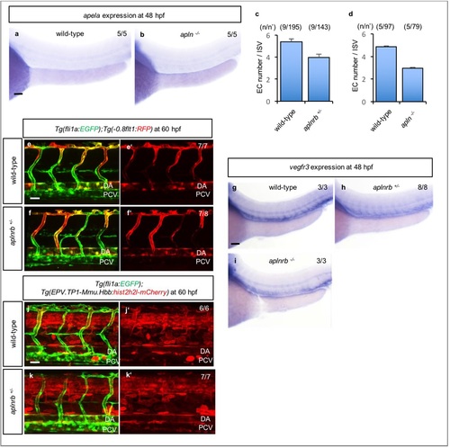
apln mutants show reduced endothelial cell numbers. (a-b) in situ hybridization of 48 hpf wild-type (a) and apln -/- (b) embryos for apela expression. The numbers of embryos examined are indicated in the top right corner of the images. (c, d) Quantitative analysis of EC numbers per three somites in 72 hpf aplnrb+/- (c) and apln -/- (d) larvae. The numbers of larvae (n) and ECs (n′) are indicated above the graph. (e-f′) Confocal images (lateral views) of 60 hpf Tg(fli1a:EGFP); Tg(-0.8flt1:RFP) wild-type (e, e′) and aplnrb+/- (f, f′) embryos. (g-i) in situ hybridization of 48 hpf wild-type (g), aplnrb +/- (h) and aplnrb -/- (i) embryos for vegfr3 expression. (j-k′) Confocal images (lateral views) of 60 hpf Tg(fli1a:EGFP); Tg(EPV.TP1-Mmu.Hbb:hist2h2l97 mCherry) wild-type (j, j′) and aplnrb+/- (k, k′) embryos. The numbers of embryos examined are in the top right corner of the images (e′, f′, g-i, j′, k′). Anterior to t 98 he left, dorsal to the top. Scale bars, 60 µm (a-b, g-i) , 20 µm (e-f′, j-k′). DA, dorsal aorta; PCV, posterior cardinal vein. Error bars, SEM. |
Expression Data
| Genes: | |
|---|---|
| Fish: | |
| Anatomical Terms: | |
| Stage Range: | Long-pec to Pec-fin |
Expression Detail
Antibody Labeling
Phenotype Data
| Fish: | |
|---|---|
| Observed In: | |
| Stage: | Protruding-mouth |
Phenotype Detail
Acknowledgments
This image is the copyrighted work of the attributed author or publisher, and
ZFIN has permission only to display this image to its users.
Additional permissions should be obtained from the applicable author or publisher of the image.
Full text @ Nat. Commun.