
Fig. S8
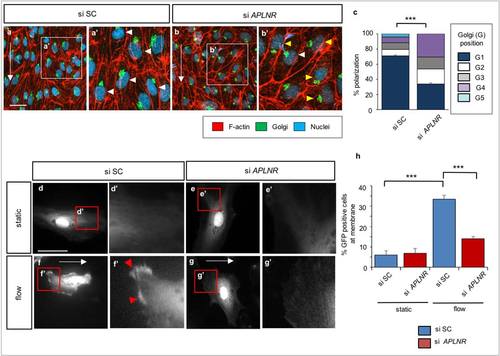
APLNR signaling modulates the polarization of human arterial endothelial cells. (a-b′) Immunofluorescence staining of HUAECs transfected with si SC or si APLNR subjected to laminar flow at 20 dynes/cm2 for 18 hours. Cells were fixed and stained with the GM130 Golgi antibody (green), Phalloidin (red) and DAPI (blue). White arrowheads point to polarized ECs (i.e., Golgi apparatus positioned within ±45° against the direction of flow (G1)). Yellow arrowheads point to depolarized ECs (i.e., Golgi positioned within + 45° to +180° and +180° to -45° against the direction of flow (G2-G4)). (c) Quantification of polarization of HUAECs subjected to laminar flow at 20 dynes/cm2 for 18 hours transfected with si SC or si APLNR. n > 300 cells, from at least three independent experiments. (d-g′) Fluorescent images of HUAECs transfected with ARRB-GFP as well as si SC or si APLNR subjected to laminar flow at 20 dynes/com2 for 15 mins. Red arrowheads point to plasma membrane localization of ARRB-GFP after 15 mins of laminar flow. (h) Quantification of ARRB-GFP membrane localization of HUAECs subjected to static condition or laminar flow at 20 dynes/cm2 for 15 mins transfected with si SC or si APLNR. *** P<0.05. Scale bars, 20 µm. Error bars, SEM. |