
FIGURE
Fig. S1
Fig. S1
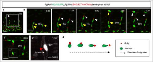
Endothelial cell polarization during migration. (a-c′) 3D-rendered confocal stack time-lapse images of the trunk region of a 36 hpf Tg(kdrl:NLS-EGFP);Tg(fli1a:B4GALT1-mCherry) embryo. (b) Time-lapse images of the white box in (a). Time (hours:mins) is shown in the top right corner of the images. White arrowheads point to polarized ECs, yellow arrowheads to non-polarized ECs. (c) A confocal stack image of the white box in (a) at t=13:03. (c′) Enlarged white box from (c). White arrow points to the lumen of the vessel. Red arrowhead points to the localization of the Golgi apparatus on the luminal side of the blood vessel. (d) Schematic representation of endothelial polarization during migration. Scale bars, 15 µm (a-c). Anterior to the left, dorsal to the top. DA, dorsal aorta; ISV, intersegmental vessel. |
Expression Data
Expression Detail
Antibody Labeling
Phenotype Data
Phenotype Detail
Acknowledgments
This image is the copyrighted work of the attributed author or publisher, and
ZFIN has permission only to display this image to its users.
Additional permissions should be obtained from the applicable author or publisher of the image.
Full text @ Nat. Commun.