
FIGURE
Fig. S5
- ID
- ZDB-FIG-160524-29
- Publication
- Ohnmacht et al., 2016 - Spinal motor neurons are regenerated after mechanical lesion and genetic ablation in larval zebrafish
- Other Figures
- All Figure Page
- Back to All Figure Page
Fig. S5
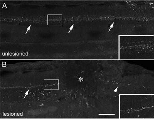
Descending TH1+ axons are present near a spinal lesion site. (Lateral views of the trunk region are shown, rostral is left; dorsal is up.) A: TH1+ axons (arrows) project down the spinal cord in the unlesioned spinal cord at 78 hpf. B: In the lesioned spinal cord, shown at 6 hours after the lesion (lesion at 72 hpf, analysis at 78 hpf), descending axons (arrow) are present rostral to the lesion site (asterisk), but have degenerated caudal to it (arrowhead). Scale bar = 75 µm and 40 µm for insets. |
Expression Data
Expression Detail
Antibody Labeling
Phenotype Data
Phenotype Detail
Acknowledgments
This image is the copyrighted work of the attributed author or publisher, and
ZFIN has permission only to display this image to its users.
Additional permissions should be obtained from the applicable author or publisher of the image.
Full text @ Development