
FIGURE
Fig. S4
- ID
- ZDB-FIG-160524-28
- Publication
- Ohnmacht et al., 2016 - Spinal motor neurons are regenerated after mechanical lesion and genetic ablation in larval zebrafish
- Other Figures
- All Figure Page
- Back to All Figure Page
Fig. S4
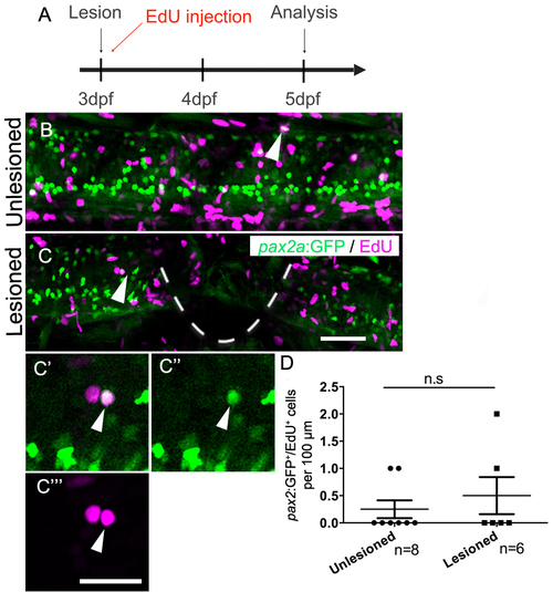
Generation of pax2a:GFP+ interneurons is not affected by a spinal lesion. (Lateral views are shown; rostral is left, dorsal is up. The lesion site is indicated by a dashed line). A: The timeline of the experiment is shown. B,C: EdU+/pax2a:GFP+ cells (arrows) are present in unlesioned (B) and lesioned (C) larvae. C′-C′′′: Higher magnifications of the cell indicated C is shown in a single optical section. D: Quantification shows no difference in pax2a:GFP+ neuron generation between 5 and 7 dpf (Mann-Whitney U test; P = 0.6839). Scale bar in C = 50 µm for B,C and in C′′′ = 20 µm for C′-C′′′. |
Expression Data
Expression Detail
Antibody Labeling
Phenotype Data
Phenotype Detail
Acknowledgments
This image is the copyrighted work of the attributed author or publisher, and
ZFIN has permission only to display this image to its users.
Additional permissions should be obtained from the applicable author or publisher of the image.
Full text @ Development