
Fig. 8
- ID
- ZDB-FIG-160524-25
- Publication
- Ohnmacht et al., 2016 - Spinal motor neurons are regenerated after mechanical lesion and genetic ablation in larval zebrafish
- Other Figures
- All Figure Page
- Back to All Figure Page
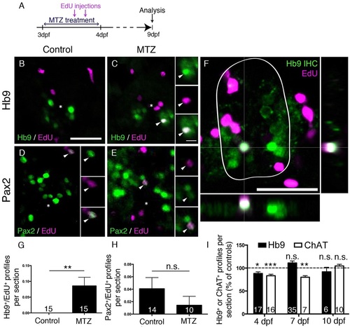
Motor neuron ablation leads to regeneration of motor neurons. (A) Experimental timeline. (B-H) In spinal cross sections, Hb9+/EdU+ motor neurons are only observed in MTZ-treated larvae (B,C, shown in a whole cross section of the spinal cord including orthogonal views in F), whereas Pax2+ interneurons are labelled by EdU in untreated and MTZ-treated larvae, quantified in G (Mann–Whitney U-test, **P=0.0063) and H (Mann–Whitney U-test, P>0.99), respectively. (I) Overall numbers of Hb9+ and ChAT+ profiles are reduced after a lesion, but return to control values at 7dpf (Hb9 only) or 10dpf (ChAT) (t-test, *P=0.0188; **P=0.0012; ***P=0.0004). Values are means±s.e.m. Scale bars: 25µm in B for B-E; 5µm in inset in C for all insets; 25µm in F. |