
FIGURE
Fig. S16
- ID
- ZDB-FIG-160425-5
- Publication
- Fu et al., 2016 - Imaging multicellular specimens with real-time optimized tiling light-sheet selective plane illumination microscopy
- Other Figures
- All Figure Page
- Back to All Figure Page
Fig. S16
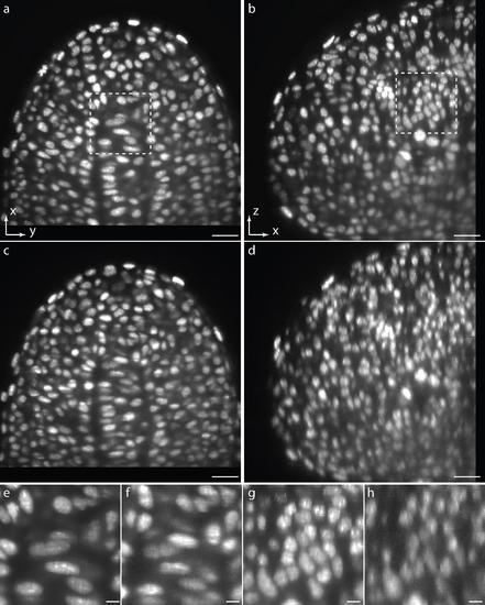
3D imaging ability comparison of regular SPIM and TLS-SPIM on a nucleus-labeled zebrafish embryo. (a, b) XY and XZ orthogonal slices of a ~15 hpf zebrafish embryo tailbud imaged by TLS-SPIM (NAOD=0.2, NAID=0.05, single excitation beam, nine tiling positions, raw image). (c, d) XY and XZ orthogonal slices of the same embryo imaged by regular SPIM with a Gaussian light sheet (NAOD=0.07, single excitation beam, raw image). (e, f) Magnified views of the selected areas in a and c. (g, h) Magnified views of the selected area in b and d. Scale bars, 20 µm (a-d), 5 µm (e-h). |
Expression Data
Expression Detail
Antibody Labeling
Phenotype Data
Phenotype Detail
Acknowledgments
This image is the copyrighted work of the attributed author or publisher, and
ZFIN has permission only to display this image to its users.
Additional permissions should be obtained from the applicable author or publisher of the image.
Full text @ Nat. Commun.