
Fig. 1
- ID
- ZDB-FIG-160425-3
- Publication
- Fu et al., 2016 - Imaging multicellular specimens with real-time optimized tiling light-sheet selective plane illumination microscopy
- Other Figures
- All Figure Page
- Back to All Figure Page
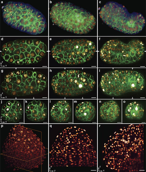
3D imaging ability of TLS-SPIM in resolving complex structures of multicellular specimens.(a-c) 3D volume renderings of a C. elegans embryo (OD95) expressing GFP::PLC∂PH (membrane) and H2B::mCherry (nucleus) at ~50 cell stage, ~200 cell stage and bean stage. (d-f) XY lateral slices through the longitudinal axis of the embryo in a, b and c. (g-i) YZ axial slices of the embryo in a-c through the positions marked in d-f. (j-o) XZ axial slices of the embryo in a-c through the positions marked in g-i. (p) 3D volume rendering of a ~15 h.p.f. zebrafish embryo tailbud expressing KikGR (nucleus). (q-r) A XY lateral slice and a XZ axial slice of the tailbud through the planes in p. Scale bars, 5 µm (d-o) and 20 µm (q-r). |