
Fig. 4
- ID
- ZDB-FIG-160316-24
- Publication
- Xu et al., 2016 - Four and a Half LIM Domains 1b (Fhl1b) Is Essential for Regulating the Liver versus Pancreas Fate Decision and for β-Cell Regeneration
- Other Figures
- All Figure Page
- Back to All Figure Page
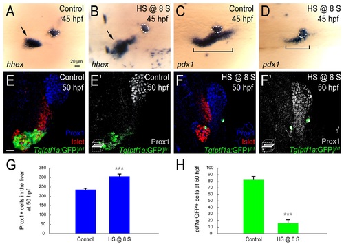
Increased Fhl1b activity suppresses specification of pancreatic cells and induces liver. (A-D) Whole-mount in situ hybridization showing the expression of hhex (A and B) and pdx1 (C and D), comparing control embryos (A and C) and fhl1b-overexpressing embryos (B and D, heat shock applied at the 8-somite stage) at 45 hpf. hhex is expressed in the liver (black arrows) and the dorsal pancreatic bud (white dotted circles). pdx1 is expressed in the developing pancreas including the dorsal pancreatic bud (white dotted circles) and intestine (black brackets), but not in the liver. When fhl1b expression was induced at the 8-somite stage, hhex expression was greatly expanded in the liver (B, black arrow), while pdx1 expression in the developing gut was reduced (D, black bracket). hhex and pdx1 expression in the dorsal pancreatic bud in fhl1b-overexpressing embryos was comparable to that of control embryos. (E-F′) Confocal images showing Islet (red), Prox1 (blue in E and F; grey in E′ and F′), and Tg(ptf1a:GFP)jh1 (green) expression at 50 hpf, comparing control embryos (E and E′) and fhl1b-overexpressing embryos (F and F′, heat shock applied at the 8-somite stage). When fhl1b expression was induced at the 8-somite stage (F and F′), the Prox1 expression domain was expanded, whereas Tg(ptf1a:GFP)jh1 expression was drastically reduced. (G) Quantification of the number (mean±SD) of Prox1-positive cells in the liver at 50 hpf. 235.5±7.3 cells were Prox1-positive in control embryos, while 306.5±12.6 cells were Prox1-positive in fhl1b-overexpressing embryos (heat shock applied at the 8-somite stage). Cells in 20 planes of confocal images from 5 individual embryos were counted. Asterisks indicate statistical significance: ***, P < 0.001. (H) Quantification of the number (mean±SD) of Tg(ptf1a:GFP)jh1-expressing cells in the exocrine pancreas at 50 hpf. The number of Tg(ptf1a:GFP)jh1-expressing cells decreased from 82.2±6.4 in control embryos to 16.0±5.2 in fhl1b-overexpressing embryos (heat shock applied at the 8-somite stage). Cells in 20 planes of confocal images from 5 individual embryos were counted. Asterisks indicate statistical significance: ***, P < 0.001. A-D, dorsal views, anterior to the left. E and F, confocal projection images; E′ and F′, confocal single-plane images, ventral views, anterior to the top. Scale bars, 20 µm. |
| Genes: | |
|---|---|
| Antibody: | |
| Fish: | |
| Condition: | |
| Anatomical Terms: | |
| Stage Range: | High-pec to Long-pec |
| Fish: | |
|---|---|
| Condition: | |
| Observed In: | |
| Stage Range: | High-pec to Long-pec |