
Fig. 2
- ID
- ZDB-FIG-160304-63
- Publication
- McPherson et al., 2016 - Motor Behavior Mediated by Continuously Generated Dopaminergic Neurons in the Zebrafish Hypothalamus Recovers after Cell Ablation
- Other Figures
- All Figure Page
- Back to All Figure Page
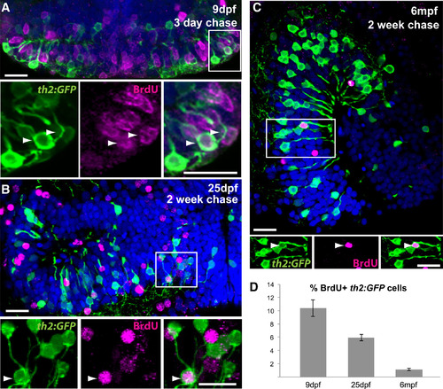
th2+ Cells Are Continuously Generated throughout Life (A) 5 dpf larvae were treated with BrdU for 24 hr and analyzed at 9 dpf. Numerous th2:GFP+ cells (green) are labeled with BrdU (magenta) in the hypothalamic posterior recess. Arrowheads indicate double-labeled cells. (B) 12 dpf larvae were treated with BrdU for 24 hr and analyzed at 25 dpf. th2:GFP+ cells (green) labeled with BrdU (magenta) can be found in medial regions of the hypothalamic posterior recess. Arrowheads indicate double-labeled cells. (C) 6 mpf fish were injected interperitoneally with BrdU and analyzed 2 weeks later. th2:GFP+ cells (green) labeled with BrdU (magenta) can be found in a midsagittal view of the hypothalamic posterior recess. Arrowheads indicate double-labeled cells. (D) Percent of BrdU+ cells in the th2:GFP+ population throughout the entire posterior recess. Error bars represent SEM; n = 5 brains. Images in (A) and (B) are ventral maximum intensity confocal z projections of the hypothalamic posterior recess. Images in (C) are maximum intensity confocal z projections from midsagittal views of the hypothalamic posterior recess. Individual and merged channels of boxed region are shown in the lower panels. The scale bars represent 10 µM. |