
Fig. 1
- ID
- ZDB-FIG-160304-62
- Publication
- McPherson et al., 2016 - Motor Behavior Mediated by Continuously Generated Dopaminergic Neurons in the Zebrafish Hypothalamus Recovers after Cell Ablation
- Other Figures
- All Figure Page
- Back to All Figure Page
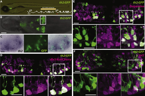
A th2:GFP Transgene Labels Dopaminergic Neurons Derived from a dlx5/6+ Precursor Lineage (A) Live zebrafish 7 dpf larva expressing th2:GFP with schematic of th2 enhancer/promoter region used for transgenes. (B) Ventral whole-mount view of th2:GFP expression in a dissected 7 dpf brain; box indicates area of posterior recess shown in (C)–(F). (C) Simultaneous in situ hybridization for th2 mRNA (left) and anti-GFP immunohistochemistry (middle) shows transcript expression in all th2:GFP+ cells (right). Ventral whole-mount view of a dissected 7 dpf brain is shown. (D) Co-expression analysis shows that most th2:GFP+ cells (green) express dlx5/6:mCherry (magenta), indicating their origin from dlx5/6+ precursors. (E) Co-expression analysis shows that most th2:GFP+ cells (green) express dopamine (magenta). (F) Co-expression analysis shows that few th2:GFP+ cells (green) express serotonin (magenta). All images in (D)–(F) are ventral maximum intensity confocal z projections of the hypothalamic posterior recess from dissected 7 dpf brains. Individual and merged channels of boxed regions are shown in the lower panels. The scale bar in (B) represents 50 µm; all other scale bars represent 10 µM. |