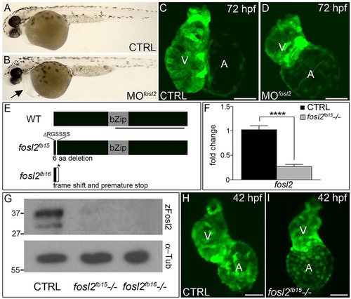
fosl2-null embryos exhibit defects in cardiogenesis. (A,B) Bright-field images of control (CTRL; A; n=44) and fosl2 morphant (MOfosl2; B; n=35) embryos at 72hpf. Morphant embryos exhibit pericardial edema (black arrow). (C,D) Confocal images of GFP+ hearts in 72hpf control (C; n=6) and fosl2 morphant (D; n=9) Tg(cmlc2:GFP) embryos. (E) Schematic diagrams of wild-type (WT) zebrafish Fosl2 with its basic leucine zipper domain (bZip) and the predicted protein products of two fosl2 mutant alleles, fosl2fb15 and fosl2fb16, generated through TALEN-mediated genome editing. The black line highlights the C-terminal region of the protein used to raise polyclonal antiserum. (F) Bar graph showing the relative levels of fosl2 mRNA in control and fosl2fb15-/- embryos at 48hpf as measured by quantitative PCR (n=6 biological replicates per group). Error bars represent s.d. ****P<0.0001. (G) Western blots of 30hpf whole-embryo lysates from control and mutant animals probed with Fosl2 (zFosl2) or α-tubulin (α-Tub) antiserum. (H,I) Confocal images of GFP+ hearts in 42hpf control (H; n=10) and mutant (I; n=4) Tg(cmlc2:GFP) embryos. V, ventricle. A, atrium. Scale bars: 50µm.
|
