
Fig. 1
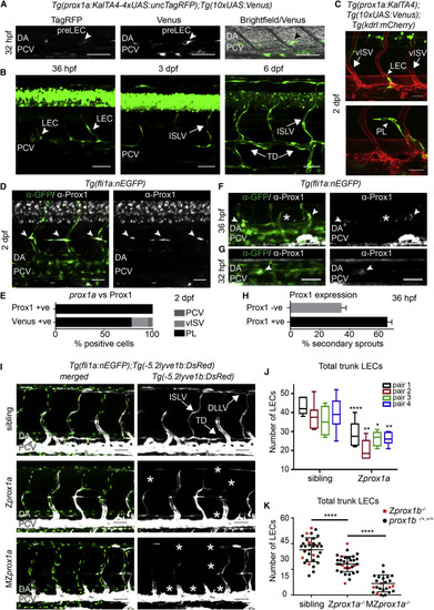
Prox1 Is Expressed and Required in Lymphatic Precursors during Secondary Sprouting (A) Single confocal projection of a TagRFP-low (left), Venus-high (middle) cell located in the wall of the PCV (bright field, right) at 32 hpf. DA, dorsal aorta; PCV, posterior cardinal vein; pre-LEC, LEC precursor (arrowheads). Scale bars, 50 µm. (B) Developmental time series of prox1a:Venus expression. PCV, posterior cardinal vein; LEC, lymphatic endothelial cell; ISLV, intersegmental lymphatic vessel; TD, thoracic duct. Scale bars, 50 µm. (C) Tg(prox1a:KALTA4);Tg(10xUAS:Venus) expression (green) in a sprouting LEC (above, arrowhead) and a parachordal lymphangioblast (PL, below, arrowhead) at 2 dpf. vISV, venous intersegmental vessel. Scale bars, 30 µm. Blood vasculature in red (Tg(kdrl:mCherry)). (D) Endogenous Prox1-positive (gray) PLs (arrowheads) in the trunk co-labeled by Tg(fli1a:nEGFP) (α-GFP, green. Scale bar, 30 µm. (E) Percentage of total prox1a:Venus- and Prox1-expressing (endogenous) cells located in the posterior cardinal vein (PCV), venous intersegmental vessels (vISV), or parachordal lymphangioblasts (PL) at 2 dpf. (F) Endogenous Prox1-positive (gray) secondary sprouts (arrowheads) and negative secondary sprouts (asterisk) at 36 hpf (Tg(fli1a:nEGFP) labeled by &alpha-GFP, green. Scale bar, 30 µm. (G) Prox1-positive (gray) nuclei located in the dorsal wall of the PCV at 32 hpf (Tg(fli1a:nEGFP) labeled by &alpha-GFP, green. Scale bar, 30 µm. (H) Mean percentage of Prox1-positive and Prox1-negative secondary sprouts per embryo at 36 hpf (Prox1 positive 65.45% ± 3.236 SEM, Prox1 negative 34.55% ± 3.236, n = 15 embryos scored across n = 6 body segments). (I) Maximum intensity projections of Tg(fli1a:nEGFP) (endothelial nuclei, green) and Tg(5.2lyve1b:DsRed) (PCV and lymphatic vessels, gray) in the trunk of sibling (top), Zprox1a (middle), and MZprox1a (bottom) mutant embryos at 4 dpf. ISLV, intersegmental lymphatic vessel; TD, thoracic duct; DLLV, dorsal longitudinal lymphatic vessel. Asterisks indicate missing lymphatic structures. Scale bar, 50 µm. (J) Box and whiskers (min to max) plot of the number of trunk lymphatic nuclei at 4 dpf in sibling and Zprox1a mutants (scored across n = 7 body segments): pair1 (n = 6 mutants, n = 10 siblings, 33% reduction), pair2 (n = 7 mutants, n = 6 siblings, 41% reduction), pair3 (n = 6 mutants, n = 4 siblings, 26% reduction), pair4 (n = 9 mutants, n = 7 siblings, 32% reduction). (K) Total number of trunk lymphatic nuclei at 4 dpf in sibling (n = 34), Zprox1a mutants (n = 32) and MZprox1a mutants (n = 24) (mean ± SEM, scored across n = 7 body segments) (t test sibling versus Zprox1ap < 0.0001 and Zprox1a versus MZprox1ap < 0.0001) (Zprox1b mutants [red], prox1b siblings [black]). |