
Fig. 2
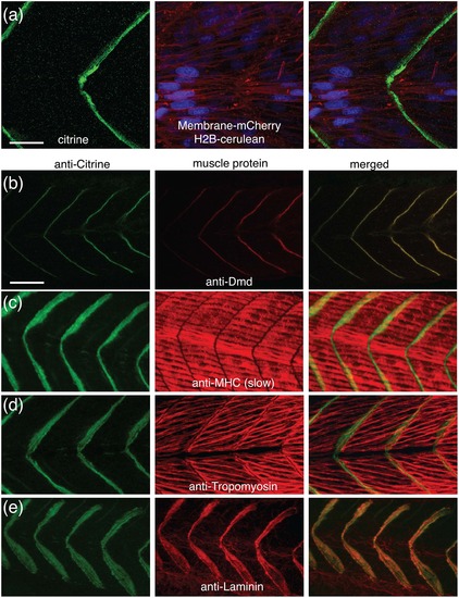
The Gt(dmd-citrine)ct90a trap line recapitulates endogenous Dmd protein expression. (a). Single optical section confocal images of live 30hpf ct90aGT embryo show that Dmd-citrine fusion protein is localized at the somite border. Dmd is in green (citrine), nuclei are in blue (H2B-cerulean), and membranes are in red (membrane-mCherry mRNA injected). (b). Antibody staining for Citrine and endogenous Dmd in the trap line confirms their co-localization at the somite border. Anti-Citrine label is in green and anti-Dmd label is in red. (c-f). Antibody staining for Citrine and endogenous muscle proteins. Myosin heavy chain (c) and Tropomyosin (d) are expressed in the muscle fibers while Laminin (e) localizes to the extracellular matrix of the myosepta. Scale bar = 10µm (a), 20µm (b). |
| Gene: | |
|---|---|
| Antibodies: | |
| Fish: | |
| Anatomical Terms: | |
| Stage: | Prim-15 |