
Fig. 4
- ID
- ZDB-FIG-150717-9
- Publication
- Stoyek et al., 2015 - Intrinsic and extrinsic innervation of the heart in zebrafish (Danio rerio)
- Other Figures
- All Figure Page
- Back to All Figure Page
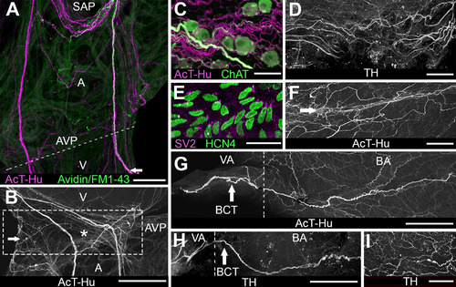
Innervation of atrioventricular region and outflow tract. A: Atrium and ventricle double labeled with AcT-Hu and neurotracer (avidin/FM1–43X, applied to right vagosympathetic trunk) show nerve trunks arising from SAP (upper part of panel) and projecting toward atrioventricular junction (dashed line; AVP: site of atrioventricular plexus). Some axons continued from AVP into ventricle (V) at lower edge of the panel. Presence of neurotracer indicates that a portion of axons originated extrinsic to the heart. B: Enlargement of region containing AVP (boxed area) from another specimen labeled with AcT-Hu shows the contribution of atrial nerve trunks to plexus; a small ganglion is indicated by the arrow; asterisk marks location of atrioventricular valve. C: Detail of AVP shows cholinergic somata associated with cholinergic and noncholinergic axons in neuropil. D: TH-labeled axons in AVP neuropil. E: HCN4-positive cells located in region of atrioventricular valve (asterisk in B). F: AcT-Hu-positive axons within atrioventricular valve leaflets (near asterisk). G: AcT-Hu-labeled innervation in walls of outflow tract (BA, bulbus arteriosus; VA, ventral aorta). Dashed line marks the border between VA and BA. A prominent nerve trunk (branchiocardiac trunk [BCT], arrow) coursed cephalocaudally in the wall of the BA toward the ventricle (right edge of panel). H: TH labeled a subset of axons in the BCT (arrow). I: BA wall was innervated with a network of fine TH-positive axons. Scale bars = 500 µm in A; 100 µm in B; 50 µm in C-E,G,I; 75 µm in F,H. |