
Fig. 5
- ID
- ZDB-FIG-150115-33
- Publication
- Fukuhara et al., 2014 - Visualizing the cell-cycle progression of endothelial cells in zebrafish
- Other Figures
- All Figure Page
- Back to All Figure Page
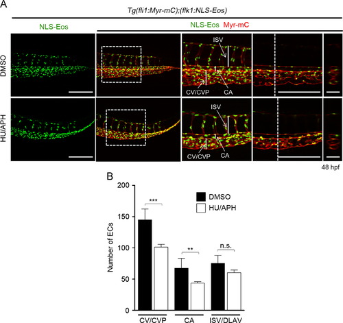
Effect of inhibiting EC proliferation on caudal vessel formation. (A) 3D-rendered confocal images of the caudal regions of 48 hpf Tg(fli1:Myr-mC);( flk1:NLS-Eos) embryos treated from 24 hpf with DMSO (upper) or HU/APH (lower). Left column, Eos images; from the second to fifth columns, the merged images of Eos (green) and mCherry (red). The boxed areas in the second column and the corresponding single scan confocal images are enlarged in the third and fourth columns, respectively. The cross-sectional single plane images of the areas indicated by dotted lines on the fourth column are also shown in the fifth column. ISV, intersegmental vessel; CA, caudal artery; CV, caudal vein; CVP, caudal vein plexus. Scale bars, 100 μm (first and fourth columns) and 50 μm (fifth column). (B) The numbers of ECs in the CV/ CVP, CA and ISV/DLAV within the five ISVs, as observed in A, were counted and shown as means±s.d. (DMSO [n=10], HU/APH [n=12]). NNp<0.01, NNNp<0.001. n.s., no significance. |
| Genes: | |
|---|---|
| Fish: | |
| Condition: | |
| Anatomical Terms: | |
| Stage: | Long-pec |
| Fish: | |
|---|---|
| Condition: | |
| Observed In: | |
| Stage: | Long-pec |
Reprinted from Developmental Biology, 393(1), Fukuhara, S., Zhang, J., Yuge, S., Ando, K., Wakayama, Y., Sakaue-Sawano, A., Miyawaki, A., Mochizuki, N., Visualizing the cell-cycle progression of endothelial cells in zebrafish, 10-23, Copyright (2014) with permission from Elsevier. Full text @ Dev. Biol.