
Fig. 4
- ID
- ZDB-FIG-141006-17
- Publication
- Moshal et al., 2011 - Discriminating Different Cancer Cells Using a Zebrafish in Vivo Assay
- Other Figures
- All Figure Page
- Back to All Figure Page
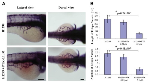
VEGF receptor tyrosine kinase inhibition mitigated tumor angiogenesis in embryonic zebrafish. Cells were stained with CM-DiI before implantation using microinjector. Immediately, after H1299 cancer cell implantation, the embryos were systemically exposed to 0.1 μM PTK 787, pharmacological inhibitor of VEGF receptor tyrosine kinase through 3 dpf. Shown are the representative whole-mount alkaline phosphatase (AP) staining of the zebrafish embryos implanted with H1299 cancer cells, (A); Quantification of number and length of the ectopic vessel originating from the developing SIV in H1299 cancer cell implanted embryos with or without PTK787 incubation (B). The error bars represent ± SEM. p values were determined by the Student′s t-test. * (p < 0.01) indicate statistically significant difference. n represents the number of embryos per group, H1299 (n = 30); H1299+PTK/0.02 μM (n = 32); H1299+PTK/0.1 μM (n = 29). Pixel (pxl) corresponds to 0.82 μm. Scale bar = 100 μm. |