
FIGURE
Fig. S6
- ID
- ZDB-FIG-140701-25
- Publication
- Wada et al., 2014 - Development of the lateral line canal system through a bone remodeling process in zebrafish
- Other Figures
- All Figure Page
- Back to All Figure Page
Fig. S6
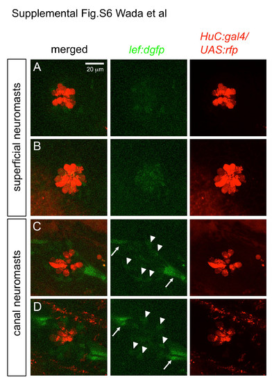
Wnt reporter activity is detectable in canal neuromasts, but not in superficial neuromasts in lef:dgfp line (13-15 mm SL, Shimizu et al., 2012). (A, B) Superficial neuromasts. (C, D) Canal neuromasts. Hair cells were visualized in the HuC:gal4; UAS:rfp transgenic lines (Kimura et al., 2008). Arrowheads indicate lef:dgfp-positive cells within neuromasts. Arrows indicate interneuromast cells, that are also lef:dgfp positive. |
Expression Data
Expression Detail
Antibody Labeling
Phenotype Data
Phenotype Detail
Acknowledgments
This image is the copyrighted work of the attributed author or publisher, and
ZFIN has permission only to display this image to its users.
Additional permissions should be obtained from the applicable author or publisher of the image.
Reprinted from Developmental Biology, 392(1), Wada, H., Iwasaki, M., Kawakami, K., Development of the lateral line canal system through a bone remodeling process in zebrafish, 1-14, Copyright (2014) with permission from Elsevier. Full text @ Dev. Biol.