
Fig. 8
- ID
- ZDB-FIG-140701-19
- Publication
- Wada et al., 2014 - Development of the lateral line canal system through a bone remodeling process in zebrafish
- Other Figures
- All Figure Page
- Back to All Figure Page
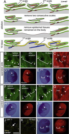
Lateral line cells are required for canal formation. (A) Schematic illustration of the lateral line cell ablation experiments. After removal of two consecutive scales (indicated by 1st and 2nd scales), the epidermal tissues remaining on the body were also removed. These epidermal tissues contain a canal neuromast and interneuromast cells. As a result, the 2nd scale regenerated in the absence of a corresponding canal neuromast. (B) Operated side. The canal epidermis (arrowheads) and a canal neuromast (stained with DiAsp) did not form within the 2nd scale (bracket). (C) The same fish shown in B was stained with Alizarin red. The 1st scale formed canal walls (arrowhead), whereas the 2nd scale could not (bracket). (D and E) The regenerated scales were removed and imaged by transmitted light microscopy. Canal walls and resorption regions were visible in the 1st scale (arrowhead in D), but not in the 2nd scale (bracket in E). Fluorescent images are shown in the right panels. Asterisk in E indicates a pore regenerated by the posterior canal epidermis. (F) Control side. Two consecutive scales (1st and 2nd) were removed. 10 days after removal, the canal epidermis was reconnected (arrowheads). (G) The same fish shown in F was stained with Alizarin red. The regenerated scales formed canal walls (arrowheads). (H and I) The regenerated scales were removed and imaged by transmitted light microscopy. Both 1st and 2nd scales formed canal walls and resorption regions (arrowheads). (J and K) The canal neuromasts did not regenerate by 10 days after scale removal in c-fms/panther fish. (L) The regenerated scale from the c-fms/panther fish also did not form canal walls or resorption regions. Asterisks indicate a pore regenerated by the posterior canal epidermis. Scale bars: 500 μm. |
Reprinted from Developmental Biology, 392(1), Wada, H., Iwasaki, M., Kawakami, K., Development of the lateral line canal system through a bone remodeling process in zebrafish, 1-14, Copyright (2014) with permission from Elsevier. Full text @ Dev. Biol.