
Fig. 6
- ID
- ZDB-FIG-140701-17
- Publication
- Wada et al., 2014 - Development of the lateral line canal system through a bone remodeling process in zebrafish
- Other Figures
- All Figure Page
- Back to All Figure Page
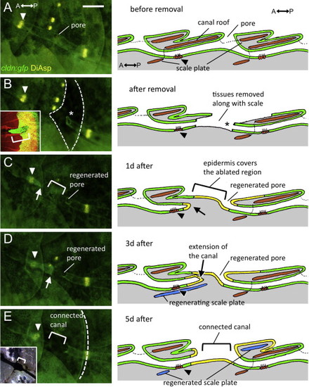
Regeneration of canal structure after removal of a lateral line scale in cldn:gfp fish. Four individuals were examined and showed the same sequence of regeneration. Representative images were taken from the same fish, with the schematic panels on the right representing the process in longitudinal section. (A) Before removal. Arrowhead indicates the canal neuromast corresponding to the scale to be removed. (B) Soon after removal. Dotted lines indicate the area where the skin epidermis was removed along with the lateral line scale. Asterisk indicates the truncated site of the posterior canal epidermis. Inset indicates the removed scale stained with Alizarin red. The posterior half of the canal epidermis was attached to the removed scale (bracket in inset), while the anterior part of canal epidermis including the canal neuromast remained on the body (B, arrowhead). (C) One day after scale removal. The new epidermis spread to heal the wound and a pore was regenerated by the truncated posterior canal epidermis (asterisk). (D) Three days after scale removal. The anterior canal epidermis extended posteriorly (arrow) to the pore region (asterisk). (E) Five days after scale removal. The extended anterior canal epidermis fused to the skin epidermis to connect the canal lumen (bracket). Caudal margin of the regenerated scale is indicated by the dotted line. Black ink was applied to visualize the regenerated canal lumen (inset). Color used in diagrams: green, epidermis; yellow, regenerated epidermis; gray, dermis; brown, lateral line scales; blue, regenerated scale. Scale bar: 100 μm. |
Reprinted from Developmental Biology, 392(1), Wada, H., Iwasaki, M., Kawakami, K., Development of the lateral line canal system through a bone remodeling process in zebrafish, 1-14, Copyright (2014) with permission from Elsevier. Full text @ Dev. Biol.