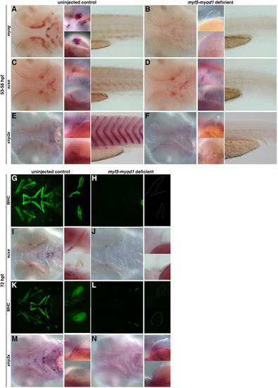
The role of muscle in the specification and maintenance of tendon populations. (A,B) Loss of myod1 and myf5 results in the complete absence of myog-positive differentiated muscles in the head (left), fin (middle) and tail (right) (93%, n=67). (C-F) In myod1-myf5-deficient embryos at 53-58hpf, scxa expression is lost in the myosepta (C,D), and xirp2a expression is completely absent in the craniofacial, pectoral fin and myoseptal tissue (E,F) compared with controls. However, loss of differentiated muscle (C,D) does not alter expression of scxa-positive tendon progenitors in the craniofacial or pectoral fin tissue (97%, n=32). (G-N) At 72hpf, myod1–/– and myf5-deficient embryos have (G,H,K,L) complete loss of myosin heavy chain (MHC) expression in the craniofacial and pectoral fin tissue and (I,J) a virtual loss of scxa expression in the head and pectoral fin tissue. (M,N) Expression of xirp2a is also missing (100%, n=19). Fluorescent images of MHC-stained flat-mounted embryos in G,H,K,L correspond to the same embryos in brightfield (I,J,M,N).
|
