
Fig. 4
- ID
- ZDB-FIG-140305-79
- Publication
- Serbedzija et al., 1998 - Regulation in the heart field of zebrafish
- Other Figures
- All Figure Page
- Back to All Figure Page
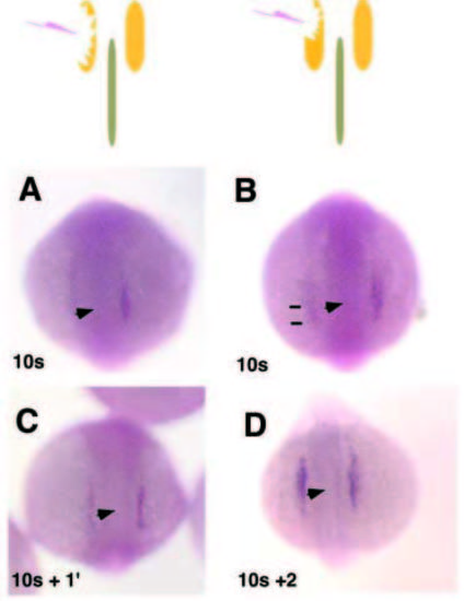
Recovery of Nkx2.5 staining after cell ablation. The schematic images indicate the location of the ablation in relation to the notochord and Nkx2.5 expression. Immediately after unilateral cell ablation in the 10-somite embryo of either (A) all of the Nkx2.5- expressing cells or (B) all of the heart progenitors; remaining posterior Nkx2.5 (marked by bars); there is no Nkx2.5 expression in the ablated region. (C) 1 hour after the ablation, the Nkx pattern of expression reappears, although the level of expression is lower than on the unablated side. (D) By 2 hours after the ablation, Nkx2.5 expression is normal. |