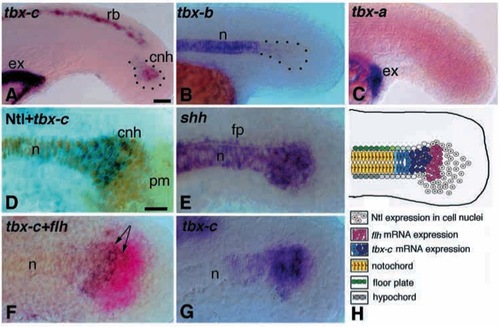
Expression pattern of tbx-c, tbx-b and tbx-a in the tail and discrete cell groups in the chordoneural hinge (CNH) of zebrafish at 18-20 hpf. (A) tbx-c is expressed in the RB cells, the excretory system and the CNH (outlined). (B) tbx-b is expressed in the notochord. (C) tbx-a is expressed in the excretory system. (D) Double staining for Ntl protein (brown) and tbx-c mRNA (blue). Ntl is expressed in the posterior mesoderm, the CNH and the notochord. tbx-c transcripts map to the anterior CNH. (E) shh is expressed in the CNH, the notochord and the floor plate. (F) Two-colour in situ hybridization staining for tbx-c (blue) and flh (magenta). tbx-c is expressed in the anterior CNH, whereas flh is expressed in the posterior part of the CNH. Expression domains of flh and tbx-c overlap in the mid-CNH (arrows). (G) tbx-c is expressed in the anterior part of the CNH. (H) A model summarizing the distribution of gene products in the CNH and the notochord. cnh, chordoneural hinge; ex, excretory system; fp, floor plate; n, notochord; pm, posterior mesoderm. Scale bars, 25 μm
|
