
Fig. 5
- ID
- ZDB-FIG-140214-25
- Publication
- Dheen et al., 1999 - Zebrafish tbx-c functions during formation of midline structures
- Other Figures
- All Figure Page
- Back to All Figure Page
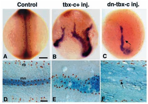
Double localization of the ntl and the Islet-1 reveals concurrent changes in the notochord and MNs of tbx-c+ and dn-tbx-c embryos at 11 hpf. The notochord is detected by ntl expression (blue) and neurons by the expression of Islet-1 (brown). (A,D) Control embryos featuring two bilateral lines of MNs along the midline and Rohon-Beard cells along the lateral neural plate. (B) Multiple notochords and extranumerary Islet-1-positive neurons in an embryo injected with a high dose of tbx-c+ mRNA. (C) Reduced notochord and a decreased number of MNs (arrow) in the dn-tbx-c embryo. (E) Extranumerary MNs with an expanded notochord in the tbx-c+ embryos. (F) The dn-tbx-c embryo showing a reduced number of MNs (arrow) and undetectable notochord. (A-C) Whole-mount embryos; (D-F) dorsal view of flat-mount embryos. mn, motor neurons; n, notochord; rb, Rohon-Beard cells. Scale bars, 100 μm (A-C); 25 μm (D-F). |