
Fig. S1
- ID
- ZDB-FIG-140211-19
- Publication
- Dyer et al., 2014 - A bi-modal function of Wnt signalling directs an FGF activity gradient to spatially regulate neuronal differentiation in the midbrain
- Other Figures
- All Figure Page
- Back to All Figure Page
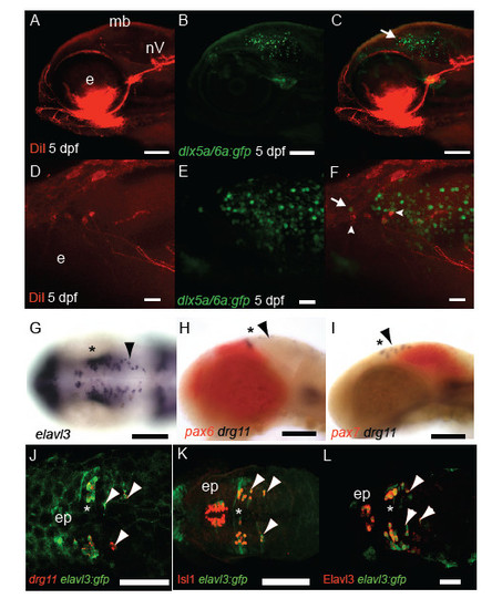
Characterisation of MTN neuronal development. DiI labelling (A,D) of jaw muscles and GFP (B,E) in Tg[dlx5a/6a:egfp] 5 dpf larvae reveals that a proportion of MTN neurons (arrowheads) co-express GFP and are larger than adjacent tectal GFP+ neurons (arrow, C,F). At 24 hpf many huc /elavl3 expressing progenitor cells are present along the A-P extent of the midbrain (G). In contrast drg11 expressing MTN (arrowheads) are restricted to the pax7 expressing midbrain (I) in comparison to nTPC neurons present in the pax6+ forebrain (H). In Tg[elavl3:gfp] embryos at 24 hpf MTN and nTPC neurons are GFP+ and express drg11 (J), Isl1 (K) and HuC protein (L). Scale bars 100 μm (A-C,G-K) 50μm (L) 20μm (D-F). |