
Fig. 1
- ID
- ZDB-FIG-140211-12
- Publication
- Dyer et al., 2014 - A bi-modal function of Wnt signalling directs an FGF activity gradient to spatially regulate neuronal differentiation in the midbrain
- Other Figures
- All Figure Page
- Back to All Figure Page
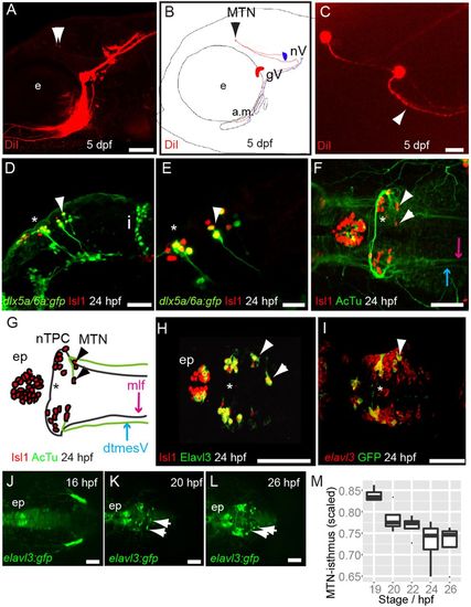
MTN neurons are the first differentiating neurons in the dorsal midbrain and innervate jaw muscles. Lateral view (A) and schematic (B) of 5 dpf zebrafish larvae with DiI (red) applied to the a.m. muscle showing labelled axons and MTN neurons (arrowheads) in the midbrain. Dorsal view (C) of DiI-labelled MTN neurons reveals a single axonal process that splits into two branches (arrowhead). Lateral views of 24 hpf Tg[dlx5a/6a:egfp] embryos with nTPC (asterisk) and MTN (arrowheads) labelled by GFP and Isl1 (D,E). Dorsal view and schematic of 24 hpf embryos with anti-Isl1 and acetylated tubulin (AcTu) labelling reveal that MTN neurons (arrowheads) pioneer the dtmesV (blue) and that nTPC neurons (asterisk) contribute to the mlf (purple) axon tracts (F,G). Dorsal views of 24 hpf embryos reveal that MTN and nTPC neurons are positive for Isl1 and Elavl3 (HuC) and express GFP in a Tg[elavl3:gfp] transgenic line (H,I); by contrast, more posteriorly located elavl3-expressing progenitor cells do not express GFP at similar stages (I). Dorsal views of Tg[elavl3:gfp] embryos at various stages reveal that MTN neurons (arrowheads) arise at the dorsal midline in the anterior midbrain and pioneer axon tracts ventrolaterally (J-L). A plot of the distance between MTN neurons and the isthmus, corrected for midbrain size, reveals that MTN neurons are formed at progressively posterior positions over time (M). gV, trigeminal ganglia; e, eye; a.m., adductor mandibulae; nV, trigeminal motoneurons; i, isthmus; mlf, medial longitudinal fasicle; ep, epiphysis; MTN, mesencephalic trigeminal nucleus; nTPC, nucleus of the tract of the posterior commissure; dtmesV, dorsal tract of the mesencephalic trigeminal. Scale bars: 100 μm in A,D,F-L; 20 μm in C,E. |