
FIGURE
Fig. 1
- ID
- ZDB-FIG-140204-45
- Publication
- Westhoff et al., 2013 - Development of an Automated Imaging Pipeline for the Analysis of the Zebrafish Larval Kidney
- Other Figures
- All Figure Page
- Back to All Figure Page
Fig. 1
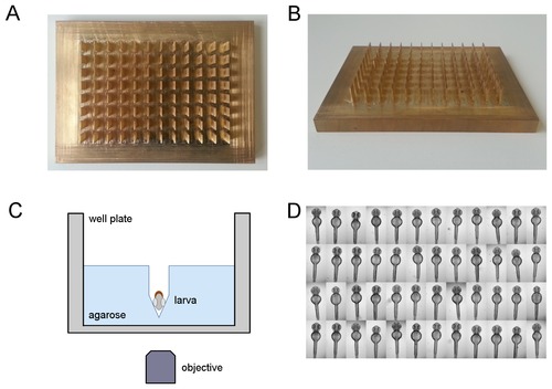
Standardized orientation of zebrafish embryos. (A,B) Photographs of the brass tool for the simultaneous generation of agarose grooves within 96 well microtiter plates: (A) top view and (B) tilted view. For dimensions of the plate see Materials and Methods section. (C) Schematic depiction of a single well with a ventrally oriented embryo within an agarose cavity. Drawing is not to scale. (D) Illustrative example of aligned and oriented embryos. Shown are dorsal views of 48 hpf embryos acquired using a 2.5x objective on an inverted wide field screening microscope. |
Expression Data
Expression Detail
Antibody Labeling
Phenotype Data
Phenotype Detail
Acknowledgments
This image is the copyrighted work of the attributed author or publisher, and
ZFIN has permission only to display this image to its users.
Additional permissions should be obtained from the applicable author or publisher of the image.
Full text @ PLoS One