
Fig. 5
- ID
- ZDB-FIG-131220-31
- Publication
- Yasuda et al., 2013 - A cis-acting element in the coding region of cyclin B1 mRNA couples subcellular localization to translational timing
- Other Figures
- All Figure Page
- Back to All Figure Page
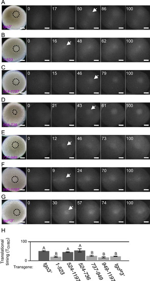
Real-time imaging of temporally regulated translation. ((A)–(G)) Real-time imaging of temporally regulated translation of tgo3′ (A), 1–523 (B), 524–1197 (C), 524–736 (D), 737–949 (E), 949–1197 (F) and tgoM32 mRNAs (G). The times after MIH stimulation are shown as standardized time TGVBD. Arrows indicate ReAsH signals detected at the first time after MIH stimulation. Dotted circles indicate the GV. Bars, 100 µm. (H) Translational timings of the reporter mRNAs after MIH stimulation. Error bars indicate mean±s.e.m. (n=3 for 1–523, 524–736 and tgoM32 n=5 for 949–1197; n=6 for 524–1197; n=7 for tgo3′ and 737–949). Transgenes indicated by B are translated earlier than those indicated by A, with statistically significant difference (P<0.05, Student2s t-test). |
Reprinted from Developmental Biology, 382(2), Yasuda, K., Kotani, T., and Yamashita, M., A cis-acting element in the coding region of cyclin B1 mRNA couples subcellular localization to translational timing, 517-29, Copyright (2013) with permission from Elsevier. Full text @ Dev. Biol.